40 light microscope with labels
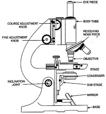
Compound Microscope Parts - Labeled Diagram and their Functions The eyepiece (or ocular lens) is the lens part at the top of a microscope that the viewer looks through. The standard eyepiece has a magnification of 10x. You may exchange with an optional eyepiece ranging from 5x - 30x. [In this figure] The structure inside an eyepiece. The current design of the eyepiece is no longer a single convex lens. Student's Guide: How to Use a Light Microscope An ultraviolet microscope uses UV light to view specimens at a resolution that isn't possible with the common brightfield microscope. It utilizes UV optics, light sources, as well as cameras. Because of the shorter wavelengths of UV light (180-400 nm), the image produced is clearer and more distinct at a magnification approximately double what ...
Compound Microscope Parts, Functions, and Labeled Diagram Compound Microscope Definitions for Labels. Eyepiece (ocular lens) with or without Pointer: The part that is looked through at the top of the compound microscope. Eyepieces typically have a magnification between 5x & 30x. Monocular or Binocular Head: Structural support that holds & connects the eyepieces to the objective lenses.

Light microscope with labels
Labeling the Parts of the Microscope Labeling the Parts of the Microscope This activity has been designed for use in homes and schools. Each microscope layout (both blank and the version with answers) are available as PDF downloads. You can view a more in-depth review of each part of the microscope here. Download the Label the Parts of the Microscope PDF printable version here. Light microscopes - Cell structure - Edexcel - BBC Bitesize Microscopes are used to produce magnified images. There are two main types of microscope: light microscopes are used to study living cells and for regular use when relatively low magnification and... Labelled Diagram Of A Light Microscope | Products & Suppliers ... Products/Services for Labelled Diagram Of A Light Microscope Microscopes - (705 companies) ...and transmission electron microscopes. Acoustic and ultrasonic microscopes use sound waves to create images of the sample. Compound microscopes use a single light path. These types of microscopes can have a single eyepiece (monocular) or a dual eyepiece...
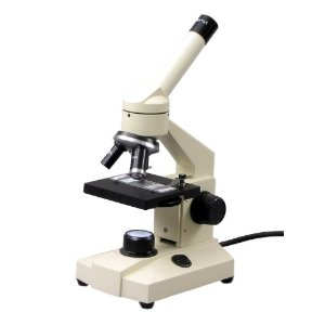
Light microscope with labels. Light Microscope - an overview | ScienceDirect Topics 6.5 Microscopy. The light microscope is an important tool in the study of microorganisms, particularly for identification purposes. The compound light microscope uses visible light to directly illuminate specimens in a two-lens system, resulting in the illuminated specimen appearing dark against a bright background. Light Microscope- Definition, Principle, Types, Parts, Labeled Diagram ... A light microscope is a biology laboratory instrument or tool, that uses visible light to detect and magnify very small objects and enlarge them. They use lenses to focus light on the specimen, magnifying it thus producing an image. The specimen is normally placed close to the microscopic lens. Microscope labels Flashcards | Quizlet Laurxyala Microscope label Terms in this set (14) ocular lens / eyepiece diopter adjustment Arm Coarse focus Fine focus On/off switch Base light source iris diaphragm Condenser Stage slide holder Objective lens Nose piece 10 terms Centrifuge Skills Test 14 terms Laurxyala Abbreviation 1 VTA 170 instruments Laurxyala Compound Light Microscope Labeling - Printable About this Worksheet. This is a free printable worksheet in PDF format and holds a printable version of the quiz Compound Light Microscope Labeling. By printing out this quiz and taking it with pen and paper creates for a good variation to only playing it online.
Microscope, Microscope Parts, Labeled Diagram, and Functions Majority of high quality microscopes used in laboratory include an Abbe condenser with an iris diaphragm. When iris diaphragm is combined with Abbe condenser, it control both the quantity of light applied as well as focus on the specimen. Aperture: It is the hole in the stage through which the base (transmitted) light reaches the stage. Explanation and Labelled Images - New York Microscope Company A fluorescence microscope is used to study organic and inorganic samples. Fluorescence microscopy uses fluorescence and phosphorescence to examine the structural organization, spatial distribution of samples. It is particularly used to study samples that are complex and cannot be examined under conventional transmitted-light microscope. Microscope Parts and Functions Microscope Parts and Functions With Labeled Diagram and Functions How does a Compound Microscope Work?. Before exploring microscope parts and functions, you should probably understand that the compound light microscope is more complicated than just a microscope with more than one lens.. First, the purpose of a microscope is to magnify a small object or to magnify the fine details of a larger ... Light Microscope: Functions, Parts and How to Use It The function of the light microscope is based on its ability to focus a beam of light through a very small and transparent specimen, to produce an image. The image is then passed through one or two lenses for magnification to view. The transparency of the specimen allows for easy and fast light penetration. Specimens can vary from bacteria to ...
Microscope Labeling - The Biology Corner 1) Start with scanning (the shortest objective) and only use the COARSE knob . Once it is focused… 2) Switch to low power (medium) and only use the COARSE knob . You may need to recenter your slide. Once it is focused.. 3) Switch to high power (long objective). Microscope Labeling - The Biology Corner Students label the parts of the microscope in this photo of a basic laboratory light microscope. Can be used for practice or as a quiz. Name_____ Microscope Labeling . Microscope Use: 15. When focusing a specimen, you should always start with the _____ objective. A Study of the Microscope and its Functions With a Labeled Diagram To better understand the structure and function of a microscope, we need to take a look at the labeled microscope diagrams of the compound and electron microscope. These diagrams clearly explain the functioning of the microscopes along with their respective parts. Man's curiosity has led to great inventions. The microscope is one of them. Parts of a microscope with functions and labeled diagram Microscopic illuminator - This is the microscopes light source, located at the base. It is used instead of a mirror. It captures light from an external source of a low voltage of about 100v. Condenser - These are lenses that are used to collect and focus light from the illuminator into the specimen.
OMAX Microscope 50mm Microscope Substage Mirror with 5mm Diameter Pin
Microscope Labeled Pictures, Images and Stock Photos Microscope Labeled Pictures, Images and Stock Photos View microscope labeled videos Browse 49 microscope labeled stock photos and images available, or start a new search to explore more stock photos and images. Newest results Fluorescent Imaging immunofluorescence of cancer cells growing... Microscope diagram vector illustration.
Using the Microscope
Parts of the Microscope with Labeling (also Free Printouts) Parts of the Microscope with Labeling (also Free Printouts) A microscope is one of the invaluable tools in the laboratory setting. It is used to observe things that cannot be seen by the naked eye. Table of Contents 1. Eyepiece 2. Body tube/Head 3. Turret/Nose piece 4. Objective lenses 5. Knobs (fine and coarse) 6. Stage and stage clips 7. Aperture

labeled animal cell under electron microscope 8745961 orig - Top Label Maker
Label the microscope — Science Learning Hub All microscopes share features in common. In this interactive, you can label the different parts of a microscope. Use this with the Microscope parts activity to help students identify and label the main parts of a microscope and then describe their functions. Drag and drop the text labels onto the microscope diagram.
Search in gallery
Microscope Types (with labeled diagrams) and Functions This is an advanced microscope that has specific application in viewing, observing and measuring the optical thickness and phase of completely transparent specimens and objects. A tiny interferometer is used and a specimen is placed on beam path of it. This path is split and then rejoined to create two superimposed images of the specimen in focus.
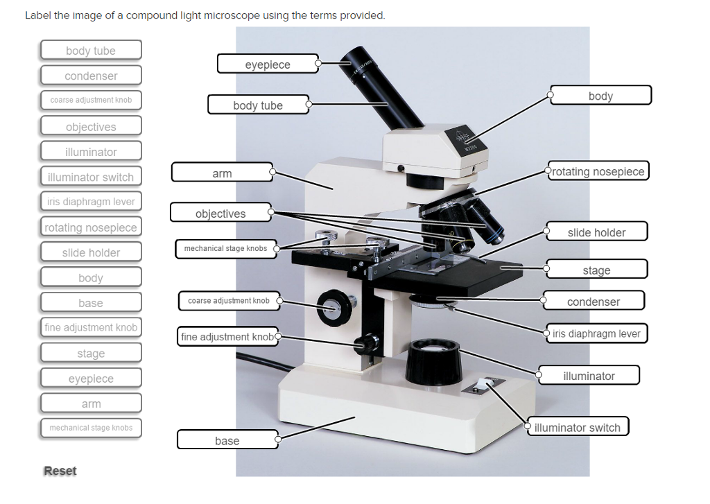
Solved: Label The Image Of A Compound Light Microscope Usi... | Chegg.com
Labeling Microscope Worksheet | Teaching Resources A straightforward worksheet in which students are required to identify the parts of a basic microscope. Tes classic free licence. Report this resource to let us know if it violates our terms and conditions. Our customer service team will review your report and will be in touch. Last updated. 21 November 2014.

Chapter 16, Page 2 - HistologyOLM 4.0
Simple Microscope - Diagram (Parts labelled), Principle, Formula and Uses A simple microscope consists of Optical parts Mechanical parts Labeled Diagram of simple microscope parts Optical parts The optical parts of a simple microscope include Lens Mirror Eyepiece Lens A simple microscope uses biconvex lens to magnify the image of a specimen under focus.

Imaging – Electronics Research Laboratory
Label a Microscope - Storyboard That Knowing the names of the different parts of the microscope is essential to be able to use one properly. Create a poster that labels the parts of a microscope and includes descriptions of what each part does. Click "Start Assignment". Use a landscape poster layout (large or small). Search for a diagram of a microscope.

Overview of a Light Microscope | Brewlab
Sperm Under Microscope with Labeled Diagram Spermatogonia under the light microscope In the germinal epithelium of a seminiferous tubule, you will find spermatogonia (stem cells) at its base. Again, the other spermatogenic cells are arranged in the order of the development process. The spermatogonia of the seminiferous tubules are immature cells that undergo several mitotic divisions.

Euglena Acus 2 - BF microscope 1250x - YouTube
Microscope Labeling Game - PurposeGames.com About this Quiz. This is an online quiz called Microscope Labeling Game. There is a printable worksheet available for download here so you can take the quiz with pen and paper. This quiz has tags. Click on the tags below to find other quizzes on the same subject. Science.
Label Light Microscope - ClipArt Best
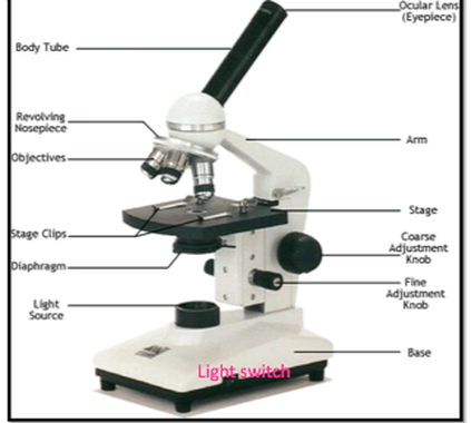
PDF Parts of the Light Microscope - Science Spot Supports the MICROSCOPE D. STAGE CLIPS HOLD the slide in place C. OBJECTIVE LENSES Magnification ranges from 10 X to 40 X F. LIGHT SOURCE Projects light UPWARDS through the diaphragm, the SPECIMEN, and the LENSES H. DIAPHRAGM Regulates the amount of LIGHT on the specimen E. STAGE Supports the SLIDE being viewed K. ARM Used to SUPPORT the

How does a microscope work? - Explain that Stuff
Labelled Diagram Of A Light Microscope | Products & Suppliers ... Products/Services for Labelled Diagram Of A Light Microscope Microscopes - (705 companies) ...and transmission electron microscopes. Acoustic and ultrasonic microscopes use sound waves to create images of the sample. Compound microscopes use a single light path. These types of microscopes can have a single eyepiece (monocular) or a dual eyepiece...
Search in gallery
Light microscopes - Cell structure - Edexcel - BBC Bitesize Microscopes are used to produce magnified images. There are two main types of microscope: light microscopes are used to study living cells and for regular use when relatively low magnification and...

Search in gallery
Labeling the Parts of the Microscope Labeling the Parts of the Microscope This activity has been designed for use in homes and schools. Each microscope layout (both blank and the version with answers) are available as PDF downloads. You can view a more in-depth review of each part of the microscope here. Download the Label the Parts of the Microscope PDF printable version here.
Microscope and its types |readbiology.com
- Labeled Microscope - Virtual Fluorescent Microscope - Wartburg College Biology Department
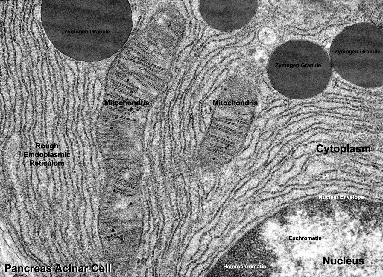
File:Pancreas acinar cell em01.jpg - Embryology
Labeling A Compound Light Microscope - ClipArt Best
Using a Light Microscope - AyushiSinhaMicroscopy
Post a Comment for "40 light microscope with labels"